Posts

Showing posts from March, 2021

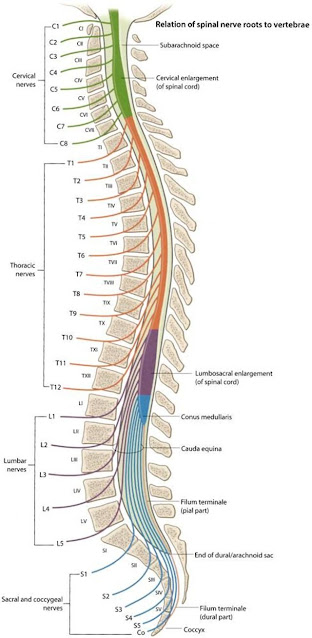
Seizure or Convulsion in pregnancy (including peripartum period)

 Seizure in pregnancy: Causes: Eclampsia - 98% (seizure due to pregnancy) Epilepsy (seizure aggravated by pregnancy) Febrile convulsion  Cerebrovascular -  Intracranial haemorrhage  Cerebral venous sinus thrombosis  Ischemic stroke  Space occupying lesion  Amniotic fluid embolism  Air embolism  Posterior reversible encephalopathy syndrome  Reversible cerebral vasoconstriction syndrome  Thrombotic thrombocytopenic purpura  Metabolic -  Hyperemesis gravidarum  Hyperglycaemia or Hypoglycaemia  Electrolyte abnormality -  Hyponatraemia, Hypernatraemia  Hypocalcaemia, Hypercalcemia Hypomagnesaemia   Pyridoxine deficiency  Acute hepatitis (due to fatty liver in pregnancy, viral hepatitis)  Uraemia  Intermittent porphyria  Infection -  Cerebral Maleria Meningitis Encephalitis Cerebral abscess  Psychogenic non epileptic seizure  Tetany  Drug withdrawal -  Cocaine  Alcohol  Local anaesthetic systemic toxicity 

Spinal cord: Cross section and tracts

Image
  The spinal cord i s 45 cm long in the adult , a measurement it shares with the lengths of the femur and the vas deferens and with the distance from the lips to the oesophagogastric junction.  It has an elongated cylindrical shape but is somewhat flattened antero - posteriorly, especially in the lumbar region . The cylinder is not uniform in diameter, but bears cervical and lumbar enlargements that correspond to the origins of the brachial and lumbosacral plexuses. The spinal cord is divided into 31 segments, each emitting a pair of spinal nerves. There are: Eight cervical segments. Twelve thoracic segments. Five lumbar segments. Five sacral segments. One coccygeal segment. With the exception of C1 and C2, the spinal nerves exit the spinal canal through the intervertebral foramina. At the terminal end of the spinal cord: The conus medullaris is the tapered terminal portion of the cord. The cauda equina is the collection of spinal nerves that continue inferiorly in the spinal canal af

Spinal cord injury: effects on respiratory system

Image
  The spinal cord is the exclusive relay of sensory, motor and autonomic information between the CNS and the peripheries. The level of spinal cord injury determines whether individual organs will remain in communication with the brain. Respiratory failure is common after spinal cord injury; respiratory complications are the most common cause of death.  With cervical and upper thoracic cord injury, the major cause of morbidity and mortality is alveolar hypoventilation combined with an inability to clear bronchial secretions.  Respiratory muscles are not affected with lumbar and low thoracic injuries, so  minimal respiratory impairment can be expected with these injuries.  The higher the spinal cord lesion, the greater the impact on ventilation: – Injury at C5 vertebral level and above Paralysis of diaphragm and all respiratory muscles.  Gross ventilatory impairment requiring immediate ventilatory support.  Usually require long-term mechanical ventilation or phrenic nerve stimulation. –

Atrial Flutter in anesthesia

  Atrial flutter is characterized by  an organized atrial rhythm with an atrial rate of 250–350 bpm with varying degrees of AV block.  Atrial flutter is a type of supraventricular tachycardia caused by a re-entry circuit within the right atrium. The length of the re-entry circuit corresponds to the size of the right atrium, resulting in a fairly predictable atrial rate of around 300 bpm (range 200-400). The rapid P waves create a sawtooth appearance on ECG and are called flutter waves.  Flutter waves are particularly noticeable in leads II, III, aVF, and V1.  The flutter waves are not separated by an isoelectric baseline.  The ventricular rate may be regular or irregular depending on the rate of conduction. Most commonly, patients have 2:1 AV conduction; an atrial rate of 300 bpm with 2:1 conduction, for example, results in a ventricular rate of 150 bpm. Characteristically the ventricular rate is about 150 bpm.  Atrial flutter frequently occurs in association with other dysrhythmias s